Lab-work · reports in progress
coursework
Lab reports and analysis code from my physics degree at NUS.
Experimental Work
hall effect in semiconductors
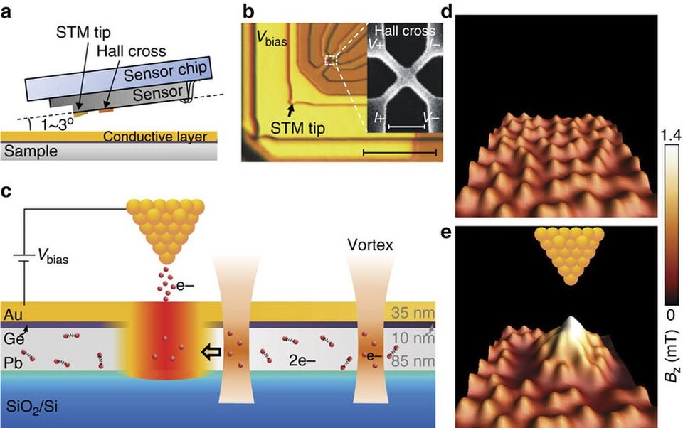
Measured Hall voltage and magnetoresistance across temperature to extract carrier type, mobility, and the intrinsic transition; includes uncertainty analysis.
raman spectroscopy
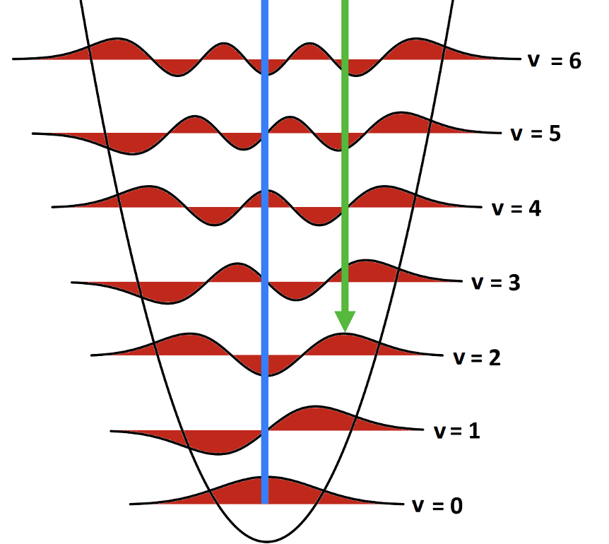
Characterised carbon allotropes via D/G band ratios, estimated graphene layer count, and identified an unknown semiconductor wafer from its 521 cm⁻¹ mode.
x-ray diffraction analysis
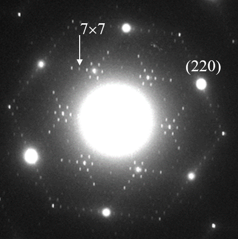
Used Bragg peaks to determine lattice constants and identify an unknown crystal; report documents calibration limits.
magnetic moment in helmholtz field
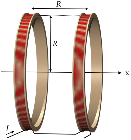
Calibrated Helmholtz field constant and tested scaling laws; corrected a model assumption during analysis.
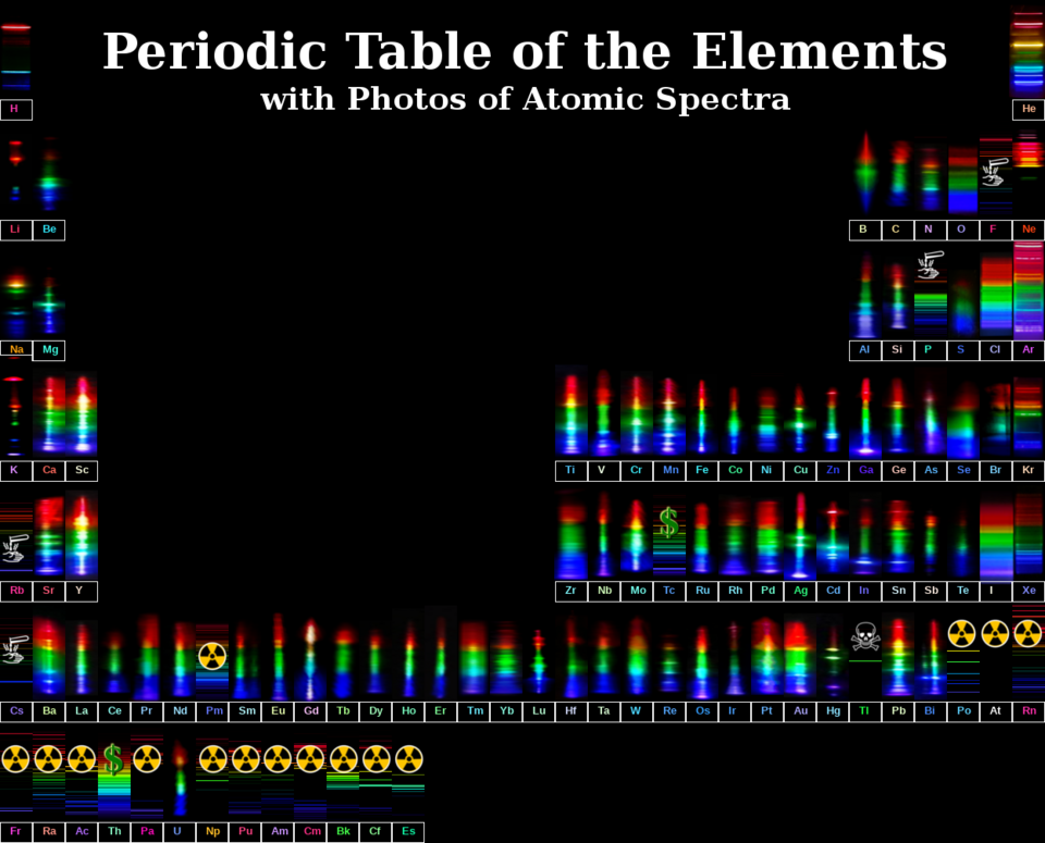
x-ray fluorescence analysis
Qualitative elemental identification of unknowns and quantitative brass composition via Gaussian deconvolution of overlapping K-lines.
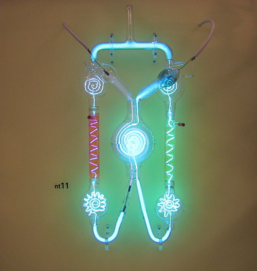
paschen curve & gas discharge
Mapped breakdown voltage vs. electrode gap at three pressures; extracted the second Townsend coefficient and identified the avalanche-to-streamer transition.
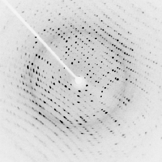
laue diffraction & x-ray crystallography
Indexed Laue back-reflection patterns and determined lattice parameters; combined with powder XRD for crystal identification.
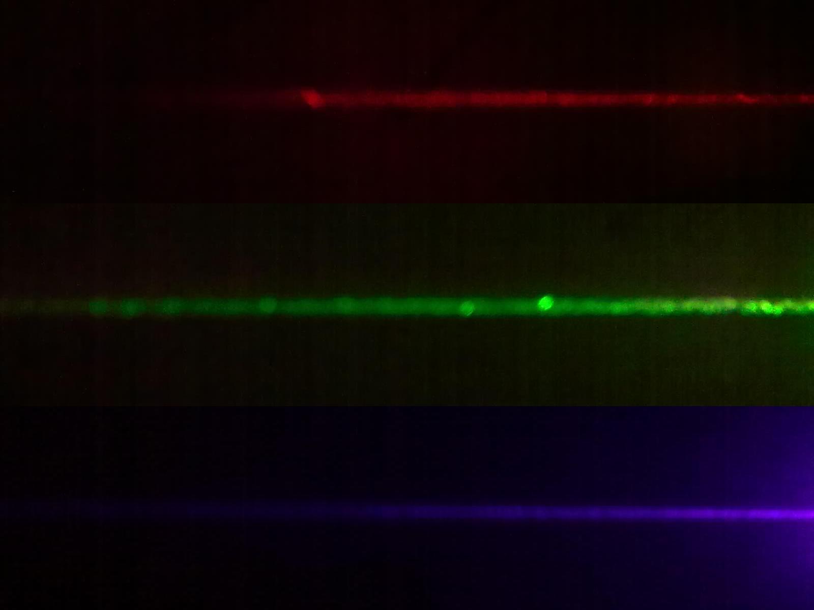
propagation of laser light (pll)
Measured Gaussian beam parameters and beam quality; report focuses on fitting and measurement limits.
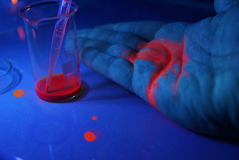
fluorescence spectroscopy: protein unfolding
Tracked acid denaturation of BSA and lysozyme via intrinsic Trp/Tyr fluorescence; observed emission shifts and intensity changes tied to tertiary structure loss.
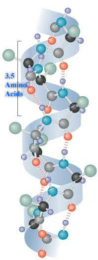
circular dichroism: protein secondary structure
Monitored α-helical content of BSA and lysozyme under acid denaturation using far-UV CD; lysozyme's disulfide bonds preserved its secondary structure.
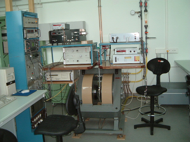
electron spin resonance (esr)
Extracted g-factor from frequency–field measurements; analyzed Zeeman splitting and systematics.
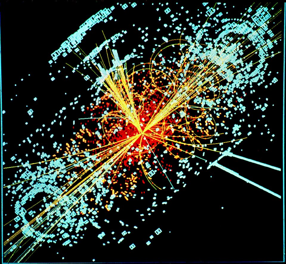
trim ion transport simulation
Monte Carlo simulations of ion range, damage, and sputtering across nuclear microscopy, semiconductor implantation, and proton therapy applications.
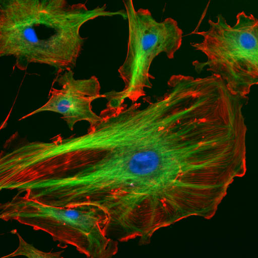
fluorescence microscopy: cellular structure
Imaged Hoechst-stained onion epidermal and cheek epithelial cells; compared brightfield and fluorescence contrast for nuclear localisation.